Root Canal – Before & Afters
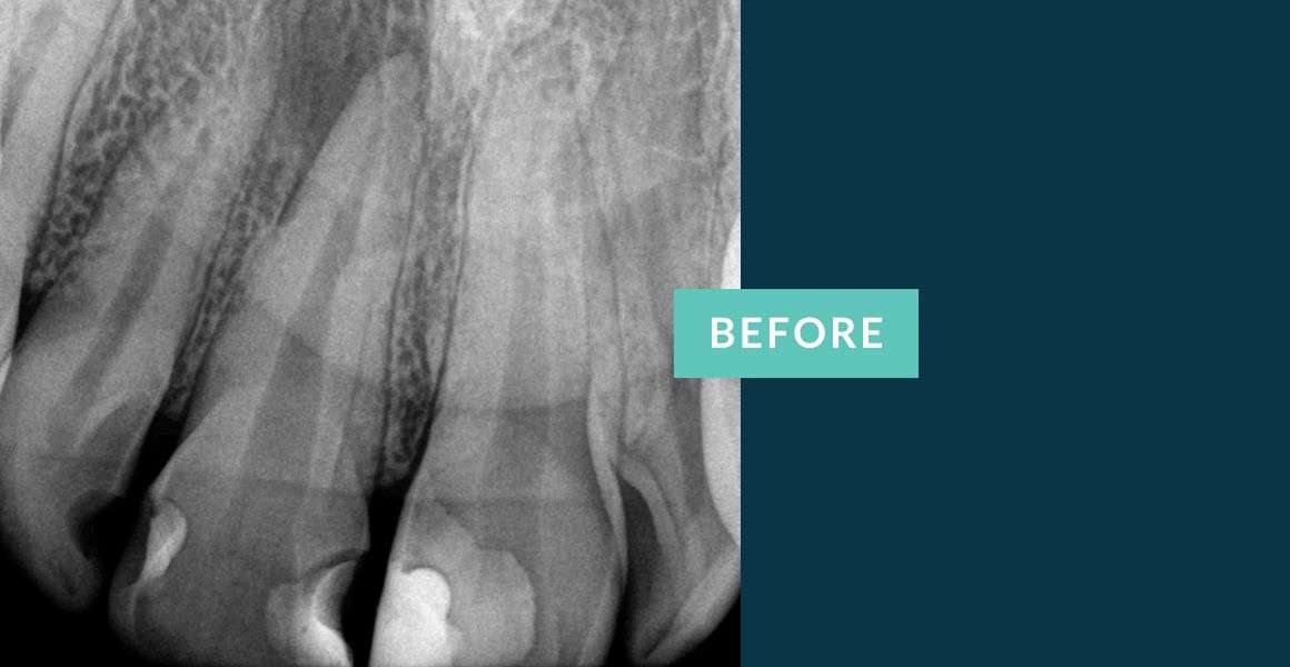
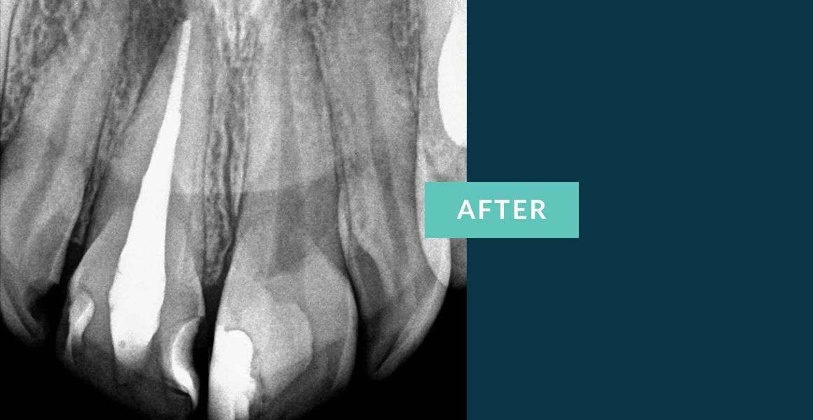
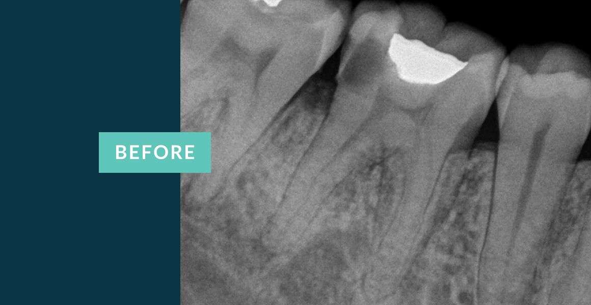
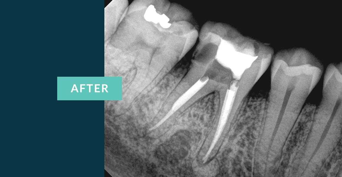
Properly treated Root Canals may stop the pain and save the tooth for decades to last. Below are just a few of the root canal before and after x-rays of endodontic treatments completed by our Root Canal Specialist, Dr. Steven Lipner.
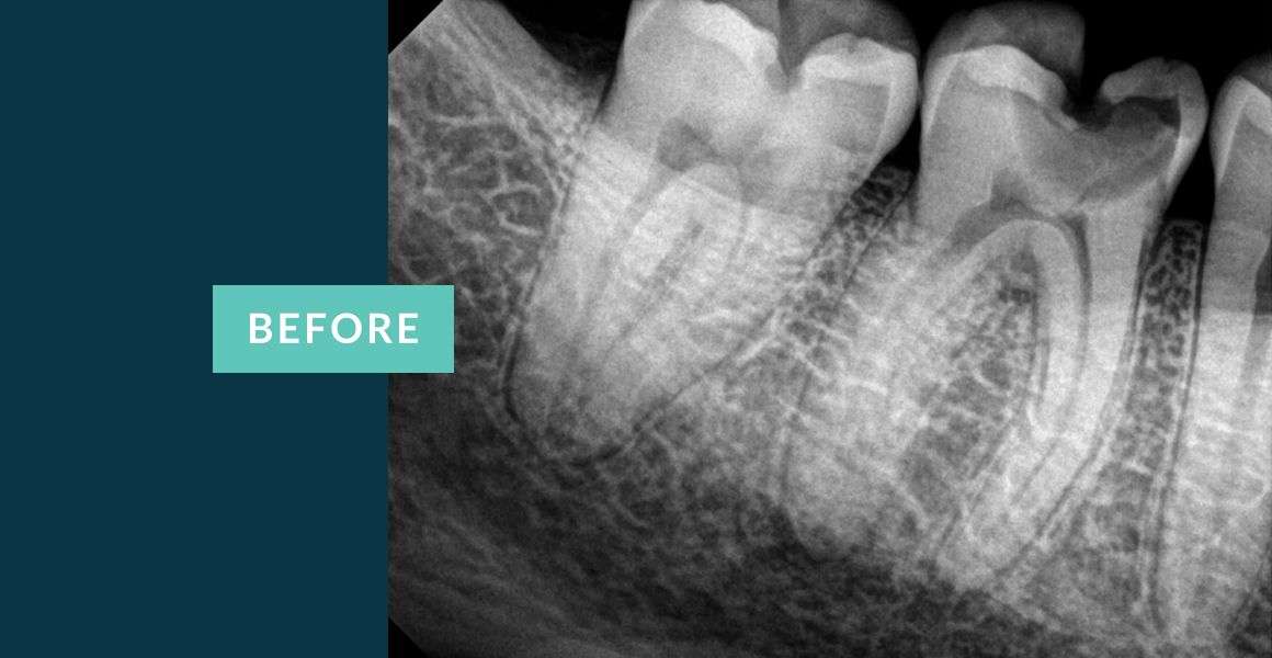
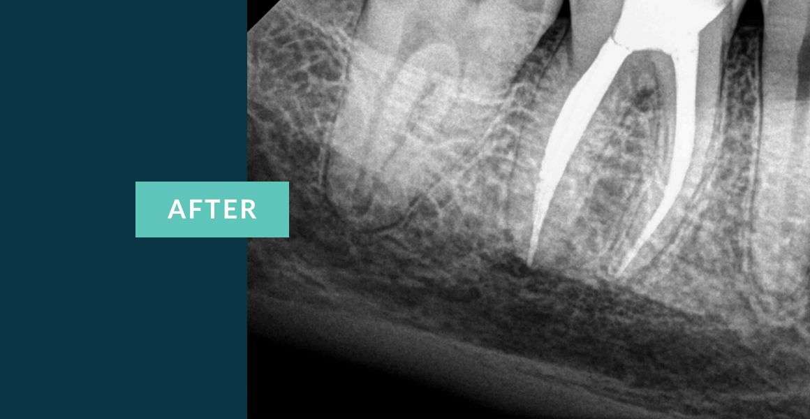
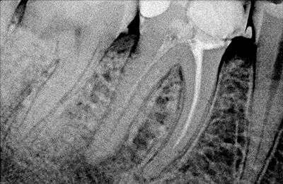
Anterior teeth typically have one canal. On the before root canal x-ray, you can see the condition of the tooth when the patient presented to the office. The reason for needing a root canal is often a recurrent decay that is too close to the nerve and/or the infection at the root of the tooth, called apex, the darker area surrounding the root, as seen on the before x-ray. After x-ray show beautifully completed root canal treatment.
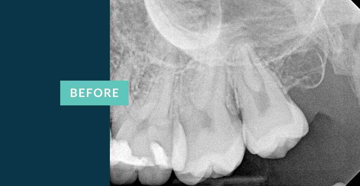
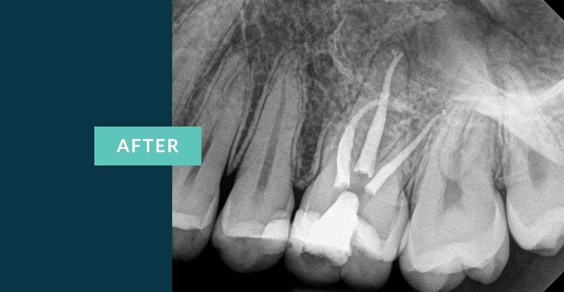
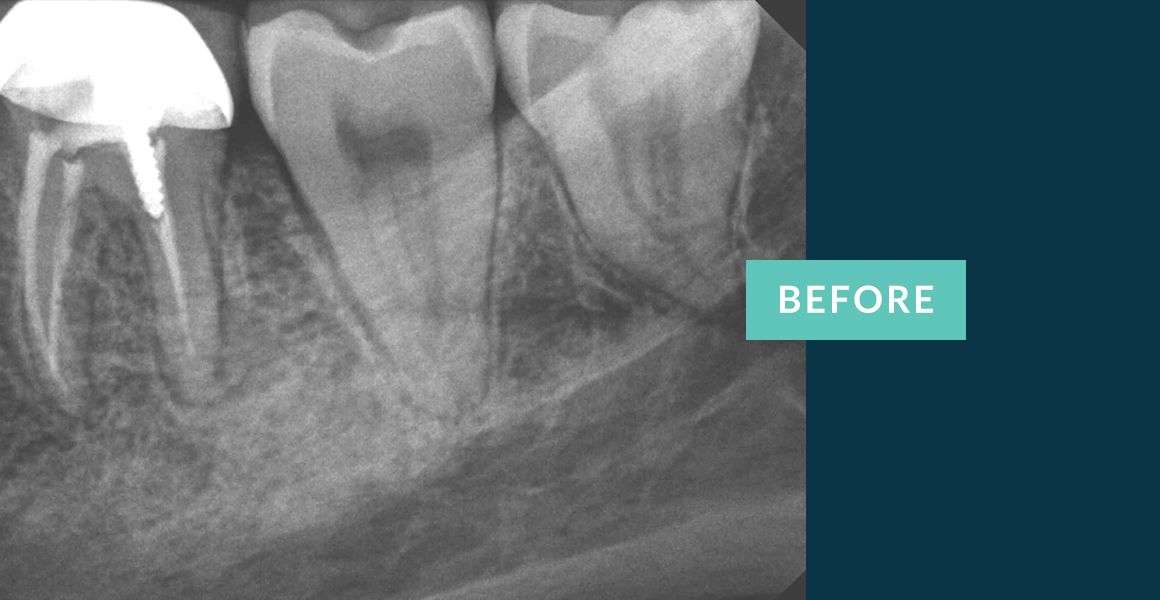
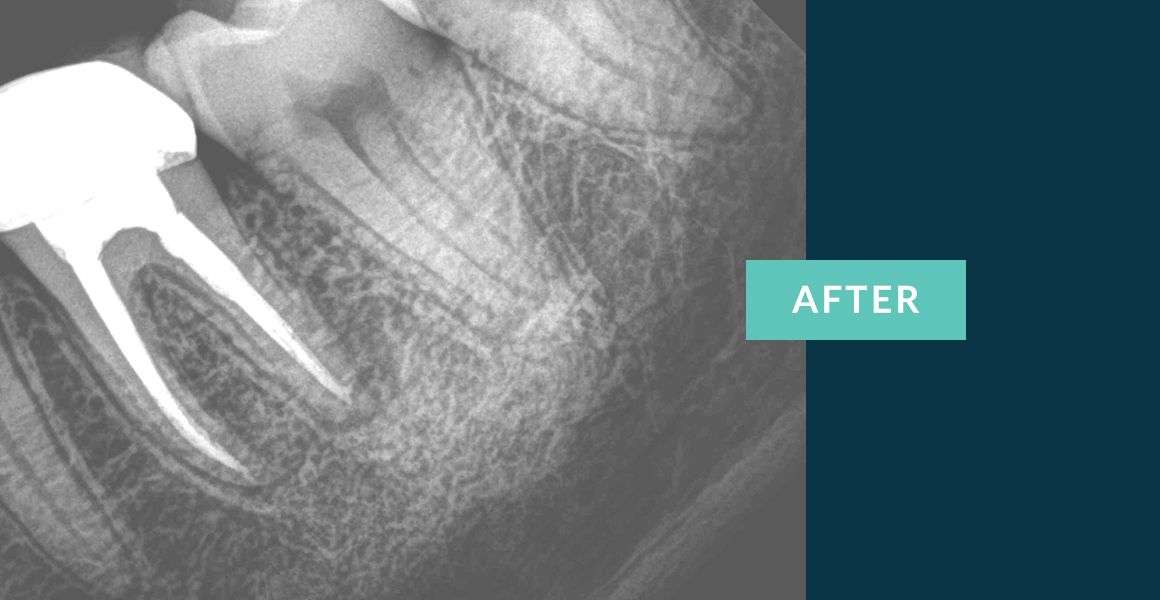
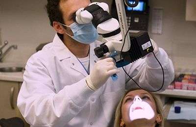
Learn MoreMolars are the largest teeth positioned in the back of the mouth. They may have 3-4 roots. These teeth are more difficult to treat because of various complications that may obstruct the procedure, like small bite, curved roots, calcified roots, tiny roots. Having proper training and expert skills is crucial to ensure proper end results. The use of an Endodontic Microscope is always beneficial to executing the best root canal on such teeth. The dark areas on the teeth on the before x-rays show decay that resulted in need of a root canal. The examples of root canals here demonstrate the scenarios mentioned above, i.e. tiny canals, curved and four canals.
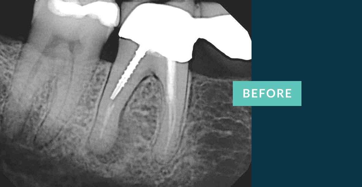
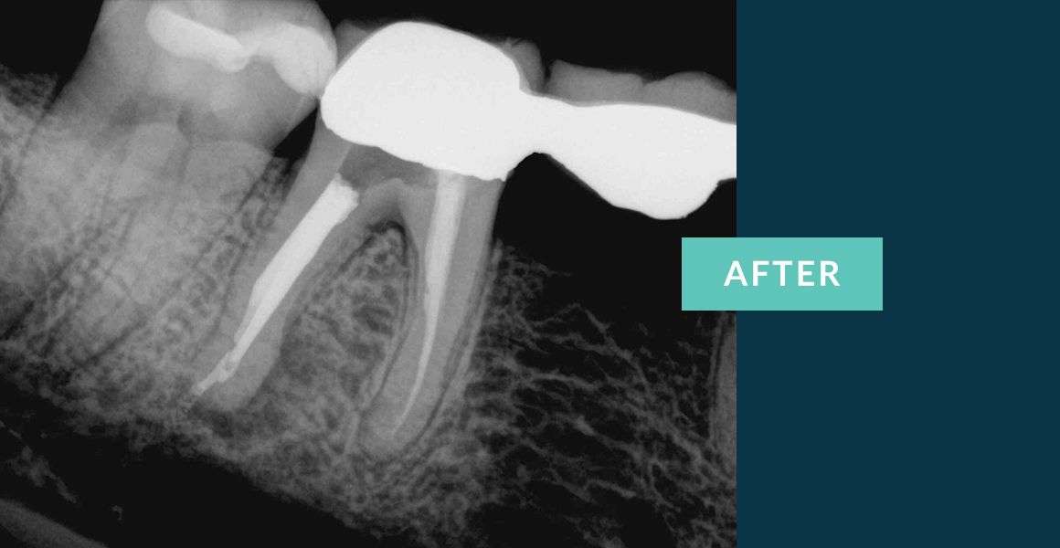
Learn MoreOften patients come to us because of the pain they are experiencing due to a failing root canal treatment. The reasons can be because one of the roots was missed. On the root canal x-ray, the roots sometimes overlap and a dentist may miss one of the roots. Another reason for missed roots can be calcified canals. Having skills, training and proper equipment is essential to saving the teeth and reversing the effects of a failing root canal. The use of the high-powered microscope helps us find those untreated and compromised roots and save the teeth from extractions.
Learn More Our Providers
 Blog
Blog
 Contact us
 Endodontics
 Root Canal Treatment
 Emergency Root Canal
 Root Canal Retreatment
 Complimentary Teeth Whitening
 Teeth Whitening
